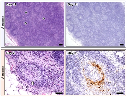
Figure 3. Lymph node changes in the 108 pfu (top) and 109 pfu (bottom) dose groups.
108 pfu dose, Day 11: Marked lymphoid hyperplasia with large numbers of secondary follicles containing well developed germinal centers (*), H&E 5X; mag bar = 100 um. Poxviral immunohistochemistry (right) is negative, anti-vaccinia IHC; 5X; mag bar = 100 um. 109 pfu dose, premature death group, Day 2: Follicular depletion and lymphocytolysis with abundant apoptotic debris, H&E 20X; mag bar = 50 um. Poxviral IHC staining (brown) is localized to the mantle zone and scattered individual cells within the paracortex and sinuses, anti-vaccinia IHC; 20X; mag bar = 50 um.

