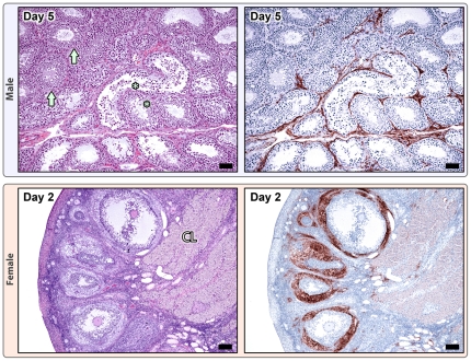
Figure 4. Gonadal changes in males (top) and females (bottom) during smallpox.
Males (108 pfu dose), Day 5: Areas of testicular (seminiferous tubule) degeneration (*) adjacent to normal seminiferous tubules (arrows), H&E 10X; mag bar = 50 um. Poxviral IHC staining (brown) is localized to the interstitium surrounding degenerate tubules; normal areas are negative, anti-vaccinia IHC; 10X; mag bar = 50 um. Females (109 pfu), Day 2: H&E shows normal ovary with primordial, primary, secondary, and tertiary follicles, and remnant corpus luteum, H&E 5X; mag bar = 100 um. Poxviral IHC staining is primarily localized to the thecal cell layer of secondary and tertiary follicles, anti-vaccinia IHC; 5X; mag bar = 100 um.

