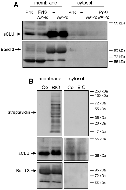
Figure 2. RBCs sCLU localizes at both extra- and intracellular sides of the plasma membrane.
(A) Representative immunoblot analysis of purified membrane and cytosol fractions of untreated RBCs (-); RBCs treated with NP-40 (NP-40) or RBCs digested with proteinase K in the absence (PrK) or presence of NP-40 (PrK/NP-40). Samples (N = 3) were probed with specific anti-sCLU and anti-Band 3 antibodies. (B) Immunobloting of streptavidin, sCLU and Band 3 in purified membrane and cytosolic fractions of control (Co) or biotinylated (BIO) RBCs (N = 3). Molecular weight markers are shown to the right of the blots.

