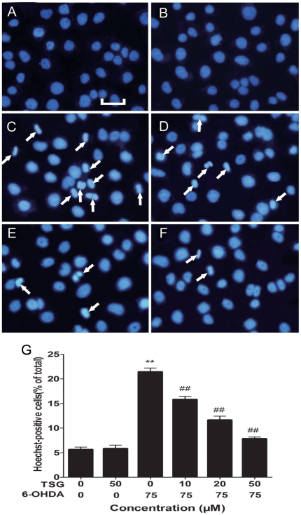
Figure 3. Fluorescence images show the nucleic changes of PC12 cells incubated in 6-OHDA with or without TSG.
Cells were stained with the DNA-binding fluorochrome Hoechst 33258. (A) shows normal culture medium nucleic morphology, (B) and (C) respectively show cells cultured in 50 µM TSG or 75 µM 6-OHDA for 24 h. In addition, cells were pretreated with 10 µM (D), 20 µM (E) or 50 µM (F) TSG for 24 h and then incubated in 6-OHDA (75 µM) for an additional 24 h. (G) Histograms showing ratio of condensed nuclei to total nuclei. White arrows represent location of apoptosis cell. Scale bars represent 50 µm. **P<0.01 versus untreated control cells; # #P<0.01 versus 6-OHDA-treated cells.

