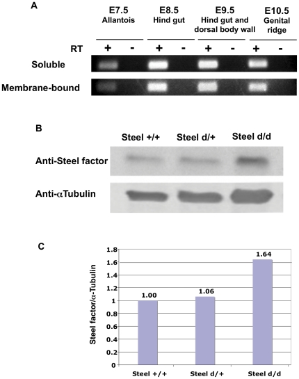
Figure 1. Expression of Steel factor in mouse embryos.
(A) RT-PCR analysis of membrane-bound and soluble Steel factor. cDNA was prepared from dissected E7.5 allantois, E8.5 hind gut, E9.5 hind gut and dorsal body wall, and E10.5 genital ridge. (B) Expression levels of soluble Steel factor protein in embryos of different Steel-dickie genotypes measured by western blot. (C) Densitometric analysis of western blots in (B). α-tubulin antibody was used as a loading control.

