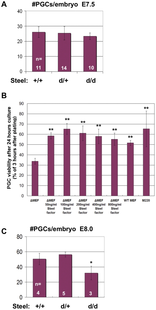
Figure 2. PGC number in Steeld/d embryos and in vitro culture.
(A) There was no significant change in PGC numbers at E7.5 in Steeld/d embryos compared to their littermates. “n” indicates the number of embryos used for quantitation. (B) PGC number after 24 hours in vitro culture in medium with or without soluble recombinant Steel factor on different feeder layer cells. Y axis represents the ratio of PGC number 24 hours after plating versus 3 hours after plating. ΔMEF: primary MEF from Steel-null embryos. M220: stromal cell line express only membrane-bound Steel factor. ** = p<0.01. (C) PGC number reduced significantly in E8.0 Steeld/d embryos compared to their littermates. “n” indicates the number of embryos used for quantitation. * = p<0.05.

