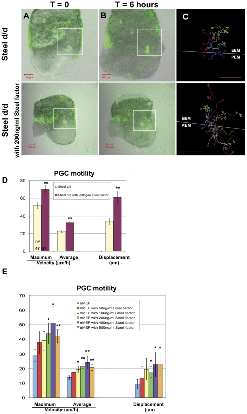
Figure 4. Effects of soluble recombinant Steel factor on PGC motility in Steeld/d embryos at E7.5 and in vitro culture.
(Column A, B) Frames at t = 0 and t = 6 hours respectively from movies of E7.5 Steeld/d embryos with or without addition of 200 ng/ml soluble recombinant Steel factor. (Column C) Tracks were made from PGCs in the allantois (white boxes) that remained in the plane of the confocal image throughout the movies. The white line indicates the boundary between the extraembryonic tissues (EEM), and the posterior end of the embryo (PEM). Scale bars in (A–C): 100 µm. (D) The maximum velocity, average velocity, and displacement of PGCs in E7.5 Steeld/d embryos were significantly increased by adding of 200 ng/ml soluble recombinant Steel factor into culture medium for 6 hours. “n” indicates the number of PGCs used for quantitation. Units on the “Y” axis vary based upon parameter, and are indicated below the bar charts. ** = p<0.01. (E) The maximum velocity, average velocity, and displacement of PGCs after 24 hours in vitro culture with increasing concentration of soluble recombinant Steel factor on Steel-null MEFs (ΔMEF). Units on the “Y” axis vary based upon parameter, and are indicated below the bar charts. * = p<0.05.

