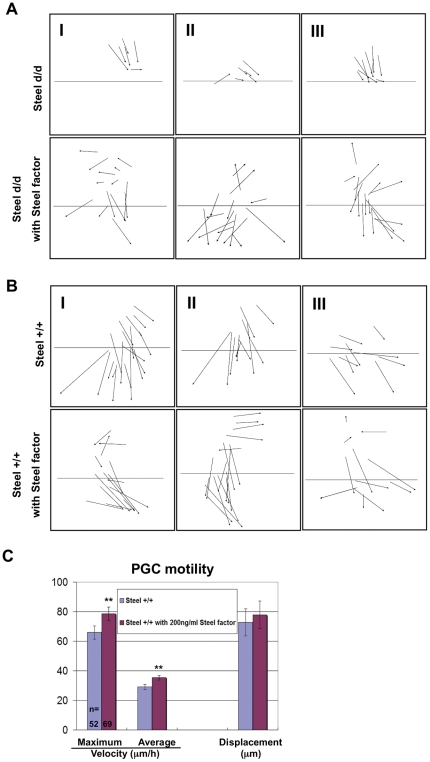
Figure 5. Effects of soluble recombinant Steel factor on PGC directions.
Directions of individual PGC migration in Steeld/d embryos (A) or wild type embryos (B) with or without addition of 200 ng/ml soluble recombinant Steel factor. The boundary between the extraembryonic tissues (EEM), and the posterior end of the embryo (PEM), is marked by a line. Column I, II, and III are representative images from 3 different embryos of the same genotype labeled on the left. (C) The maximum velocity, average velocity, and displacement of PGCs in E7.5 wild type embryos with or without addition of 200 ng/ml soluble recombinant Steel factor into culture medium for 6 hours. “n” indicates the number of PGCs used for quantitation. Units on the “Y” axis vary based upon parameter, and are indicated below the bar charts. ** = p<0.01.

