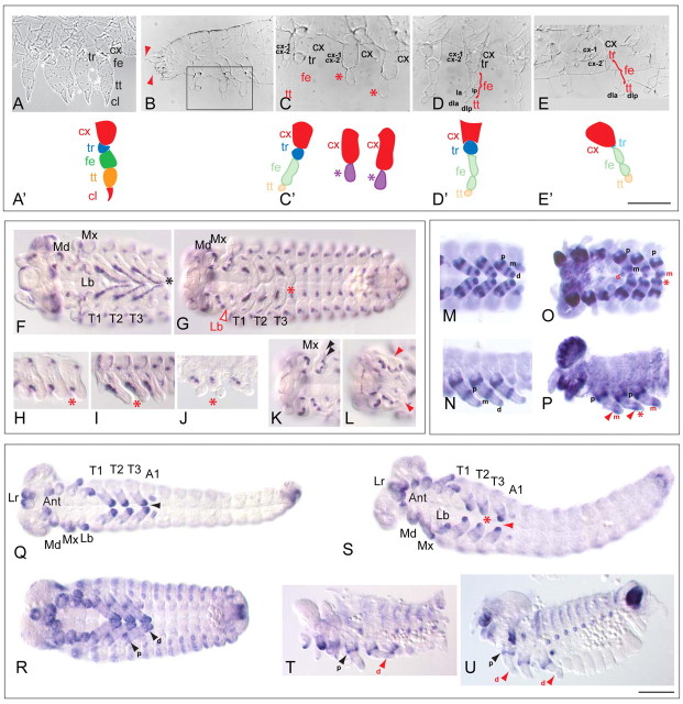
Fig. 6.
Tc-fz1 has an exclusive function in appendage differentiation. (A,A′) Wild-type larval leg showing coxa (cx), trochanter (tr), femur (fe), tibiotarsus (tt) and pretarsal claw (cl). Schematic of leg parts is shown in A′. (B-C′) Tc-fz1RNAi larva. Legs have defects of the proximal-distal axis. (B) Tc-fz1RNAi larva, anterior half. Antennae lack flagellae (red arrowheads). Boxed area in B is enlarged in C and illustrates a severe leg phenotype. Schematic is shown in C′. The coxa are unaffected according to coxa specific bristles cx-1 and cx-2 but femur and tibiotarsus are fused and twisted. There is no distinct identity of leg segments (asterisks and purple colour in C′), no pretarsal claw is present and the trochanter is fused to the coxa. (D-E′) Weaker Tc-fz1RNAi phenotypes. Schematics of leg phenotypes are shown in C′ and D′. (D,D′) Tc-fz1RNAi larva showing ‘non-pareille’ phenotype (Grossmann et al., 2009). Ectopic constriction in the femur, no pretarsal claw according to tibiotarsal bristle markers dla and dlp. Femur bristle markers la and lp are shown. (E,E′) Tc-fz1RNAi larva showing a stronger phenotype as in D. Trochanter is not clearly recognisable (blue in E′; ‘pearls on a chain’ phenotype) (Grossmann et al., 2009). (F) Tc-wg expression in wild type in all appendages, reaching in a ventral stripe to the tip of each leg (asterisk). (G) Tc-wg expression in a Tc-fz1RNAi embryo is restricted to the coxa (red asterisk), appendages are distally fused and twisted (red asterisk). There is normal Tc-wg expression in the segments. (H-J) Consecutively older Tc-fz1RNAi legs. Tc-wg expression remains proximal, restricted to coxa and never extends into the tips (asterisks). In J, the distal part of the legs shows the most severe defect (asterisks). (K) Tc-wg expression in wild-type head appendages. Maxillar lobes are present (arrowheads). (L) Tc-wg expression in Tc-fz1RNAi head appendages. Maxillar palp is missing (red arrowhead). (M,N) In wild-type legs (ventral and lateral view) Tc-Lim is expressed in three domains: the proximal ring (p, coxa), the median ring (m, femur/tibia) and the distal domain (d). (O,P) Tc-Lim expression in Tc-fz1RNAi legs, ventral and lateral view. Distal domain is missing or only partly present (red asterisks), the median ring (m) broadens (arrowheads). (Q,R) Tc-dachsous expression in wild-type appendages. In younger embryos, dachsous is expressed in a prominent distal domain in all appendages (Q), and later additionally in a proximal ring in the coxa (arrowheads in R). (S-U) Tc-dachsous expression in Tc-fz1RNAi embryos. Distal expression in the legs is incomplete or missing, only proximal expression remains. (S) Tc-fz1RNAi embryo with leg defects. The distal expression of Tc-dachsous is reduced (asterisk, arrowhead). (T,U) Older Tc-fz1RNAi legs. Only the proximal expression of Tc-dachsous together with a very faint distal domain (arrowheads). Leg segment bristle markers: cx-1, coxa-1; cx-2, coxa-2. A1, abdominal segment 1; Ant, antennae; cl, pretarsal claw; cx, coxa; d, distal ring; dla, distal dorsal anterior; dlp, distal dorsal posterior; fe, femur; la, dorsal anterior; Lb, labial segment; lp, dorsal posterior; Lr, labrum; m, median ring; Md, mandibular segment; Mx, maxillar segment; p, posterior ring; T1-T3, thoracic segments 1-3; tr, trochanter; tt, tibiotarsus. Red asterisks indicate affected structures. Scale bar: 100 μm.

