


Fig. 1.
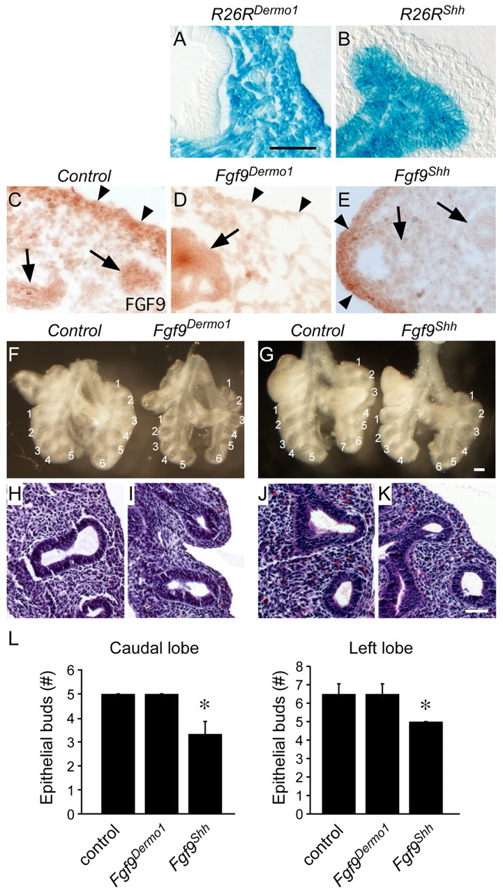
Mesothelial and epithelial FGF9 have unique functions in developing mouse lung. (A,B) lacZ staining for the Rosa26 reporter (R26R) targeted with Dermo1-Cre (A) and Shh-Cre (B) lungs at E10.5 showing mesenchymal/mesothelial and epithelial cell-specific β-gal staining, respectively. (C-E) Immunohistochemical detection of FGF9 in E12.5 lung showing expression in epithelium (arrows) and mesothelium and submesothelial mesenchyme (arrowheads) in control lungs (C). Fgf9Dermo1 lungs (D) have greatly reduced staining in submesothelial mesenchyme (arrowheads) and Fgf9Shh lungs (E) have reduced expression in epithelium (arrows). (F,G) Anterior views of gross dissections of control (Dermo1-Cre, Fgf9f/+) and Fgf9Dermo1 (F) and control (Shh-Cre, Fgf9f/+) and Fgf9Shh (G) lungs at E12.5. Note the decreased mesenchyme but normal epithelial branch number and orientation of lobes with the Fgf9Dermo1 lung (F) and decreased number of epithelial branches and normal mesenchyme in Fgf9Shh lung (G). (H-K) Hematoxylin and Eosin-stained histological sections of control (H,J), Fgf9Dermo1 (I) and Fgf9Shh (K) lungs at the same stages as shown in F,G. (L) Number of epithelial buds in the caudal and left lobes at E12.5. *P<0.001 (Student's t-test). Error bars represent s.d. Scale bars: 25 μm in A-E,H-L; 200 μm in F,G.