


Fig. 3.
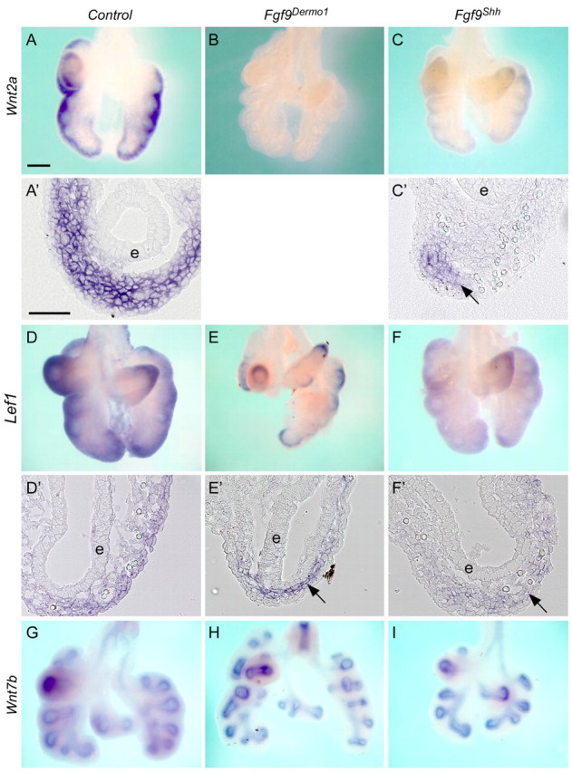
Mesothelial FGF9 regulates WNT2A expression and mesenchymal WNT/β-catenin signaling. (A-C′) Expression of Wnt2a in control (A,A′), Fgf9Dermo1 (B) and Fgf9Shh (C,C′) mouse lung mesenchyme at E12.5. A′ and C′ are cryosections of the corresponding whole mounts. Note that in Fgf9Dermo1 lung mesenchyme Wnt2a is not detected. In Fgf9Shh lung, Wnt2a is still expressed in the distal mesenchyme (arrow) but not in proximal mesenchyme of epithelium (e). (D-F′) Expression of Lef1 in control (D,D′), Fgf9Dermo1 (E,E′) and Fgf9Shh (F,F′) lung mesenchyme at E12.5. D′, E′ and F′ are cryosections of the corresponding whole mounts. Note that in Fgf9Dermo1 lung, Lef1 expression was detected in the proximal mesenchyme (arrow) and in Fgf9Shh lung Lef1 was detected in the distal mesenchyme (arrow). (G-I) Wnt7b expression in lung epithelium was not significantly changed in control (G), Fgf9Dermo1 (H) and Fgf9Shh (I) lung epithelium at E12.5. Scale bars: 200 μm in A-C,D-E,G-I; 50 μm in A′,C′,D′-F′.