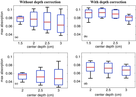
Figure 5.
The box and whisker plot of the maximum absorption of the layers in the lesion region. Homogenous low-contrast lesion of size 2 cm diam reconstructed (a) without and (b) with depth correction. Homogenous low-contrast lesion of size 3 cm diam reconstructed (c) without and (d) with depth correction.

