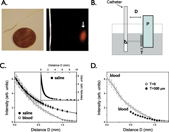
Figure 1.
Catheter prototype for intravascular sensing of NIR fluorescence signals. (a) The NIRF catheter consists of a 0.36-mm∕0.014-in. floppy radiopaque tip with a maximum outer diameter of 0.48 mm∕0.019 in.. The arrow highlights the focal spot (40±15 μm) for the 90-deg arc-sensing catheter at a distance of 2±1 mm (arrow). (b) Phantom experiment to measure NIR light attenuation in the presence of whole blood. Plaque (P) consists of 1% Intralipid plus India ink 50 ppm plus AF750 (an NIR fluorochrome, concentration 300 nmol∕L); tissue (T: fibrous cap) consists of polyester casting resin plus titanium dioxide plus India ink; a container (gray shaded area) was filled with fresh rabbit blood or saline. The catheter was immersed in fresh rabbit blood and positioned at variable distance (D) from a fluorescent phantom representing the plaque (P). To mimic the presence of a fibrous cap, a solid tissue phantom of thickness T was interposed between the plaque and the lumen. (c) Plot of detected NIRF signal as a function of distance D in presence of blood compared to saline, showing only modest attenuation by blood. Inset, fluorescence signal decay in saline at distance of up to 10 mm. (d) Plot of the detected NIRF signal in blood in the presence of a tissue phantom (T) of thickness 500 μm shows modest NIRF signal attenuation (<35%) vs the case in (c) where T=0. Reproduced by permission from Ref. 22.

