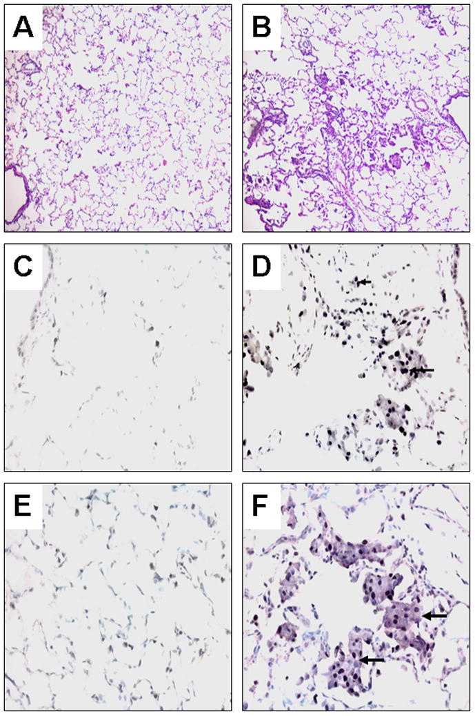
Fig. 1.

Histological examination and immunoperoxidase staining of ferret lung sections. Left panel, normal lung; Right panel, squamous metaplasia induced by tobacco smoke after 6 weeks of exposure; Hematoxylin-eosin staining at low magnification x 100 (A, B), Immunoperoxidase staining with anti-proliferating cell nuclear antigen (PCNA) antibody using 3, 3′-diaminobenzidine (DAB) plus nickel as the substrate (giving the black color) and a methyl green counterstain at high magnification x 400 (C, D), Double labeling immunoperoxidase staining with anti-cytokeratin polypeptide 19 (Vector®NovaRed as the substrate, giving the reddish-purple color) and anti-PCNA antibodies (DAB plus nickel as the substrate) and a methyl green counterstain (E, F). Arrowheads depict nuclei stained with anti-PCNA antibody. Arrows depict the cytoplasm of a cluster of cells stained with anti-cytokeratin polypeptide 19 antibody.
