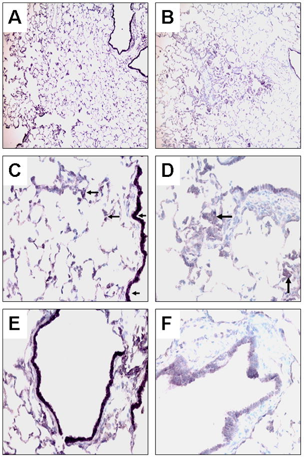
Fig. 2. Double-labeling immunoperoxidase staining of ferret lung sections with antibodies for cytokeratin polypeptide 19 (Vector®NovaRed as the substrate) and retinoic acid receptor (RAR)β (DAB plus nickel as the substrate) with methyl green counterstain.

The left panel represents normal lung sections and the right panel represents lung squamous metaplasia and adjacent bronchioles after 6 weeks of smoke exposure at low magnification x 100 (A, B) and high magnification x 400 (C –F). Arrowheads point to representative nuclei expressing RARβ (stained in black). Arrows point representative areas expressing cytokeratin polypeptide 19 (stained in reddish-purple).
