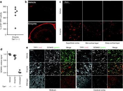
Figure 4.
Uptake of intrathecally administrated recombinant human TPP1 (rhTPP1) and effect on subunit c of mitochondrial ATP synthase (SCMAS) storage in brain. Fifteen week-old Tpp1(−/−) mice were administered three daily 80 µl infusions of artificial cerebrospinal fluid (CSF) (vehicle) (n = 4) or 10 µg/µl rhTPP1 (enzyme) (n = 6) and killed 24 hours after the final dose. (a) Relative TPP1 activity in enzyme treated animals. (b) TPP1 immunohistochemistry in cerebellum lobes IV/V. (c) TPP1 immunohistochemistry in different regions of the cerebral cortex. (d) SCMAS levels by quantitative western-blot analysis. Each symbol represents an average of multiple measurements (n ≥ 2) from individual animals. The difference between the vehicle and enzyme-treated group is significant using the Mann–Whitney nonparametric test (P < 0.01). (e) Double-label indirect immunofluorescence using the mouse monoclonal anti-human TPP1 antibody 8C4 (red in merge) and rabbit anti-SCMAS antibodies (green in merge). Note that the low signal for TPP1 in the wild type animal is due to poor immunoreactivity of 8C4 for murine TPP1 and that endogenous mitochondrial SCMAS is not detected under these conditions.45 Images were obtained using either a Nikon Eclipse E600 microscope with a ×20 objective (b) or a Zeiss LSM510 laser scanning confocal microscope with a 63x objective (c and e).

