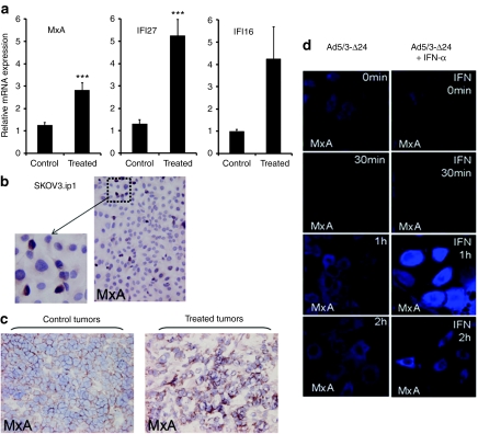
Figure 3.
Expression of Myxovirus resistance protein A (MxA) and interferon stimulated genes in tumors recurring after oncolytic adenoviral therapy. (a) RNA extracted from untreated control and Ad5/3-Δ24 treated tumors was reverse transcripted to cDNA and quantitative reverse transcriptase-PCR was performed to determine relative MxA, IFI27 and IFI16 mRNA levels (fold change). Data represent the mean ± SEM (n = 5). (b) Immunohistochemical MxA protein staining in untreated SKOV3.ip1 ovarian carcinoma cell line shows a small subpopulation of MxA positive cells. (c) Immunohistochemistry for MxA in treated tumors versus control tumors: higher MxA positive subpopulation was found in virus-resistant treated tumors. (d) MxA expression in SKOV3.ip1 cells after infection with adenovirus; To simulate the effect of stromal cells, SKOV3.ip1 cells were treated with recombinant universal type I interferon-α (IFN-α). The IFN-α treatment resulted in further upregulation of MxA expression 1 hour after infection (right panel).

