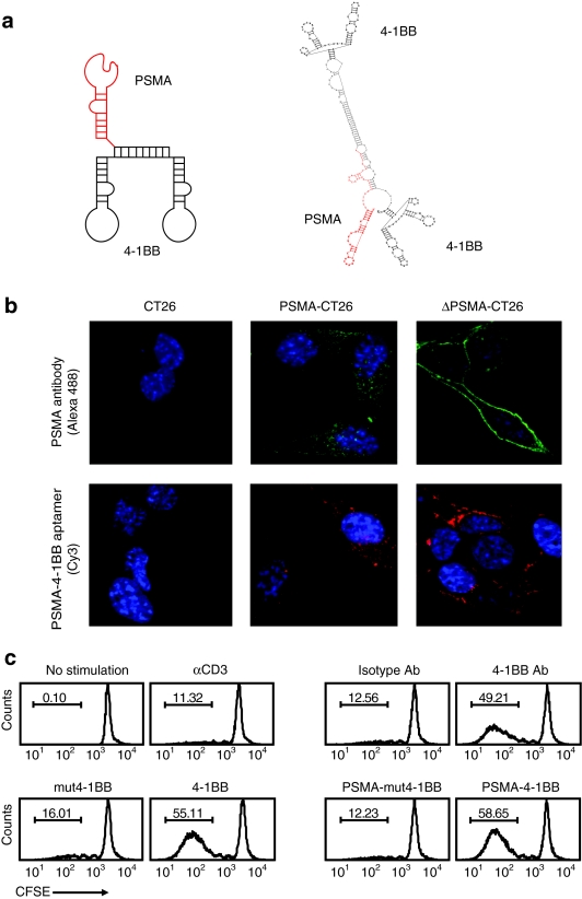
Figure 1.
Functional characterization of a bi-specific PSMA-4-1BB aptamer conjugate. (a) Sequence and computer generated secondary structure of the PSMA-4-1BB aptamer conjugate. See Methods section for full sequence. (b) Binding to PSMA-expressing CT26 tumor cells. Parental CT26 cells, CT26 cells expressing a wild-type PSMA (PSMA-CT26) or CT26 cells expressing an internalization-deficient mutant (ΔPSMA-CT26) were incubated with anti-PSMA antibody (green) or Cy3-conjugated PSMA-4-1BB aptamer conjugate (red) and analyzed by confocal microscopy (×60 magnification). Nuclei were stained with DAPI (blue) (N = 3). (c) 4-1BB costimulation. CD8+ T cells were labeled with CFSE, activated with suboptimal concentrations of anti-CD3 antibody, and incubated with anti-4-1BB antibody/isotype control IgG, unconjugated agonistic 4-1BB/costimulation-deficient mut4-1BB aptamers, or with PSMA-4-1BB /PSMA-mut4-1BB aptamer dimer conjugates. Two days later cells were analyzed by flow cytometry (N = 2). CFSE, carboxyfluorescein succinimidyl ester; PSMA, prostate specific membrane antigen.

