Figure 3.
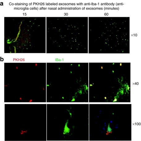
Exosomes administered intranasally are taken up by microglial cells. A total of 10 µg of PKH26-labeled EL-4 exosomes (red) were administered intranasally to C57BL/6j mice. (a) Mice were sacrificed 15, 30, and 60 minutes after intranasal administration of PKH26-labeled EL-4 exosomes. Brain tissue sections were fixed as described in the Materials and Methods section. Frozen sections (30 µm) of the anterior part of the brain were stained with the antimicroglial cell marker Iba-1 (green color). Slides were examined and photographed through an upright microscope with an attached camera (Olympus America, Center Valley, PA). Representative photographs of brain sections of mice having been intranasally administered PKH26-labeled exosomes for varying times (a) or 60 min at low (×40) and high (×100) magnification are shown (b). Each photograph is representative of three different independent experiments (n = 5). Original magnifications ×10, ×40, and ×100.

