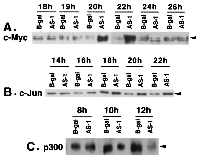
Figure 3.
Levels of c-MYC, c-JUN, and p300 in serum-starved MCF10A cells infected with AS p300 vectors. (A) Western immunoblot of c-MYC in quiescent MCF10A cells infected with AS-1 or β-gal vectors. Twenty micrograms of the protein from cell lysates prepared from MCF10A cells at indicated time points was immunoblotted by using an α-c-MYC polyclonal antibody (sc-764; Santa Cruz Biotechnology). (B) Western analysis of c-JUN in serum-starved MCF10A cells infected with Ad vectors. Twenty micrograms of protein from the cell lysates used in A was assayed by Western immunoblotting by using an α-c-JUN antibody (sc-1694; Santa Cruz Biotechnology). (C) Time course of p300 depletion in serum-starved MCF10A cells. Fifty micrograms of the protein lysates at different time periods of infection was analyzed by Western immunoblotting by using an α-p300 antibody (sc-584, Santa Cruz Biotechnology).

