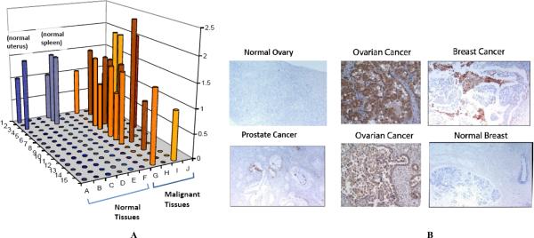
Fig. (2). uPAR expression in normal and tumor tissues.
A) Tissue microarray (TMA)data of normal and randomly selected tumor tissue. TMA FDA801 (normal tissue) and FDA801-2 (normal and tumor) were purchased form Biomax (Rockville, MD) and analyzed for uPAR expression by immunohistochemistry (IHC) using ATN-658. IHC was performed by Phenopath Laboratories and immunostaining was reviewed and scored by a board certified pathologist. B) IHC using uPAR antibody on selected tumor sections. Random tumors and normal sections were analyzed for uPAR using ATN-658 immunostaining.

