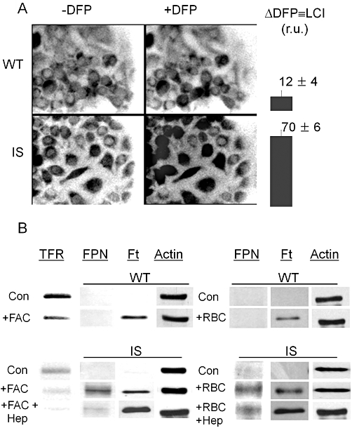
Figure 3.

Iron loading and erythrophagocytosis by RAW 264.7 [wild type (WT) and iron-sensitive (IS)] macrophages: effects on labile iron pools, ferroportin, ferritin and transferrin receptor levels. WT and IS cells were incubated 4–18 h in regular culture medium supplemented with 100 µM ferric ammonium citrate (FAC). (A) The cytosolic labile cell iron (LCI) levels in 4 h iron-loaded WT and IS cells were assessed by fluorescence microscope imaging with the fluorescent intracellular iron probe Calcein blue as described in the Methods section. LCI is shown for one experiment in relative fluorescence units determined in five different fields of cells (mean ± standard error of the mean) as determined from the increase in fluorescence 15 min after addition of 100 µM deferiprone (DFP) (Δ DFP). The range of LCI in iron loaded cells (in relative fluorescence units r.u. obtained with the same microscope settings in three independent experiments) was 4–20 for WT cells and 62–95 for IS cells. (B) Ferroportin (FPN), transferrin receptor (TfR) and ferritin (Ft) levels in WT (top panel) and IS (bottom panel) cells were assessed by Western blotting after overnight incubation of cells without or with 100 µM of FAC (+FAC) or after 1 h with or without opsonized erythrocytes (+RBC), followed by overnight incubation with or without 1 µM Hepcidin (+Hep). The data displayed are representative of one of five experiments performed in two different laboratories, showing similar Western blot patterns.
