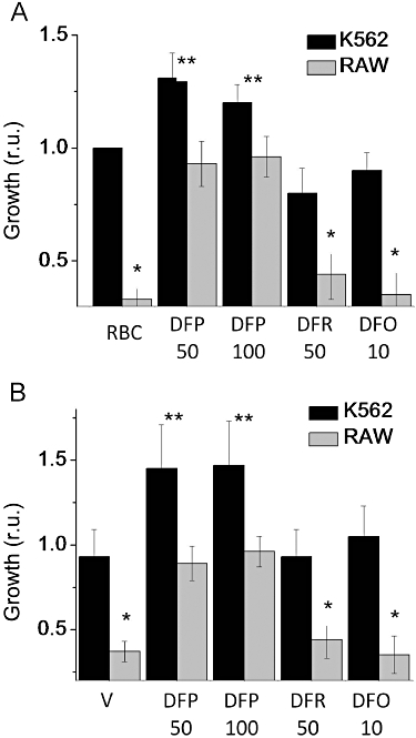
Figure 7.

Chelator-mediated iron relocation from iron-loaded RAW 264.7 macrophages to iron-depleted K562 cells in co-culture. Iron-sensitive (IS) cells were iron-loaded either (A) by incubation for 1 h with opsonized erythrocytes to allow erythrophagocytosis, followed by washing of erythropcytes and incubation overnight in culture conditions followed by removal of non-internalized RBC by hypotonic lysis and washing, or (B) by overnight exposure to 500 µM Venofer (V) and washing. Iron-deprived K562 cells were added on top of the RAW 264.7 cells and the mixed cell culture was grown for 24 h in iron-free, apo-transferrin containing medium supplemented with the iron chelators deferiprone (DFP), deferasirox (DFR) or deferrioxamine (DFO) at the indicated concentrations (µM). The K562 cells, which grow in suspension, were carefully separated from the substrate-attached RAW 264.7 IS cells by aspiration and the relative numbers of K562 cells and of RAW cells were quantified in a fluorescence plate reader following staining with the DNA fluorescent stain Hoechst 33342. Data are expressed as mean growth (in fluorescence units) relative to the control ± standard deviation (n = 3). The asterisks denote statistically significant differences between treatment and the respective control (* for RAW cells and ** for K562 cells).
