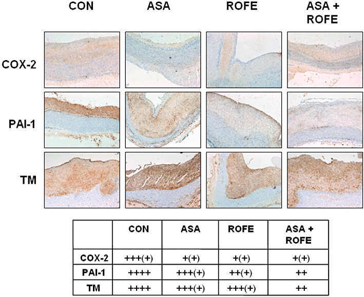
Figure 5.

Immunohistochemical staining for COX-2, TM and PAI-1 in sections of the aortic arch of rabbits fed standard diet (SD, n = 11) or atherogenic (CON, n = 13) diet for 12 weeks are shown. COX-2 expression is reduced by ROFE (n = 7), ASA (n = 8) and the combination of both (ASA + ROFE, n = 5) compared with animals fed an atherogenic diet alone. A semiquantitative scale: no staining (−), mild (+), moderate (++), strong (+++) and intensive (++++) staining was used in order to evaluate staining intensity of the sections by five independent observers in a blinded fashion (see table).
