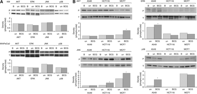
Figure 3.
Effect of supernatants on intracellular signalling proteins. Cells were cultured with BCG supernatants for 24 h before western blotting for the proteins indicated. AKT, ERK, JNK and p38 MAPK were the proteins assessed as they represented a broad range of signalling elements indicating receptor activation and intracellular functioning. Samples were designated ‘BCG’ if treated with BCG supernatants, and expressions were compared with those from tumours cultured with CONT supernatants (‘un’) and basal expressions (‘B’: where tumours were cultured in standard medium). Both the phosphorylated (P) and total (T) levels were assessed, and densitometry were performed showing the effect of supernatants on the phospho:total ratio. Cell lines that were unaffected by BCG supernatants with regard to HLA1 expression are shown in (A), while those exhibiting increases in HLA1 expression following treatment in BCG supernatant are shown in (B). *P<0.05 when compared with the untreated control as determined by paired t-tests.

