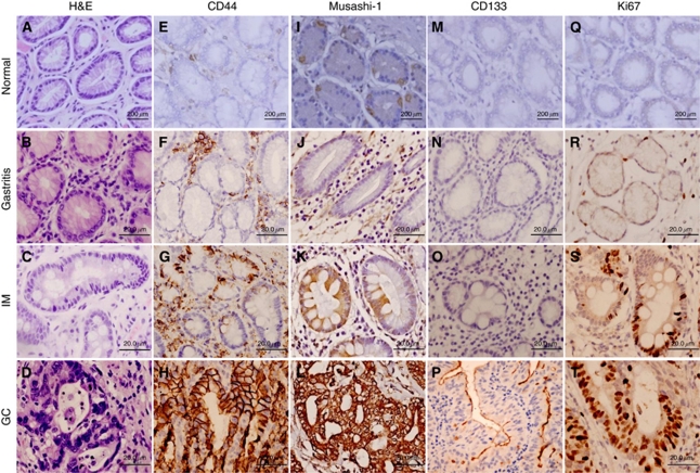
Figure 1.
Putative stem cell marker expression during the Correa pathway of gastric carcinogenesis. Representative images of normal, gastritis, intestinal metaplasia (IM) and malignant (GC) epithelial tissues from gastric specimens are shown following immunohistochemical staining for CD44 (E–H), Musashi-1 (I–L) and CD133 (M–P). Representative haematoxylin and eosin (H&E) stains (A–D) and immunohistochemical staining for Ki67 (Q–T) are also shown.

