Figure 1.
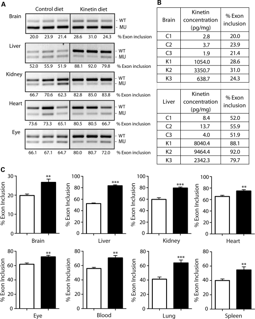
Kinetin modifies IKBKAP splicing in TgFD1 transgenic mice. (A) Representative images of RT–PCR analysis of IKBKAP exon 20 splicing in different tissues from TgFD1 transgenic mice. Expression of the FD IKBKAP mRNA produces both the WT (including exon 20) and MU (excluding exon 20). Percent exon 20 inclusion (shown below each panel) was calculated using the integrated density values (IDV) obtained for each WT and MU band. An increase in the percent exon 20 inclusion is observed in all tissues tested in the kinetin-fed mice compared with the vehicle controls. (B) Table shows the kinetin concentrations and the percent exon 20 inclusion in control (C1, C2, C3) and kinetin-fed (K1, K2, K3) mice, respectively. (C) Plotted bar charts show the quantification of the percent exon 20 inclusion in multiple tissues in kinetin-fed mice and control mice. Black and white bars represent the kinetin-fed and vehicle control mice, respectively. Error bars represent SEM (n= 8). Significant differences observed between the kinetin-fed and vehicle control mice are represented by ** (P< 0.01) and (*** P< 0.0005); Student's t-test.

