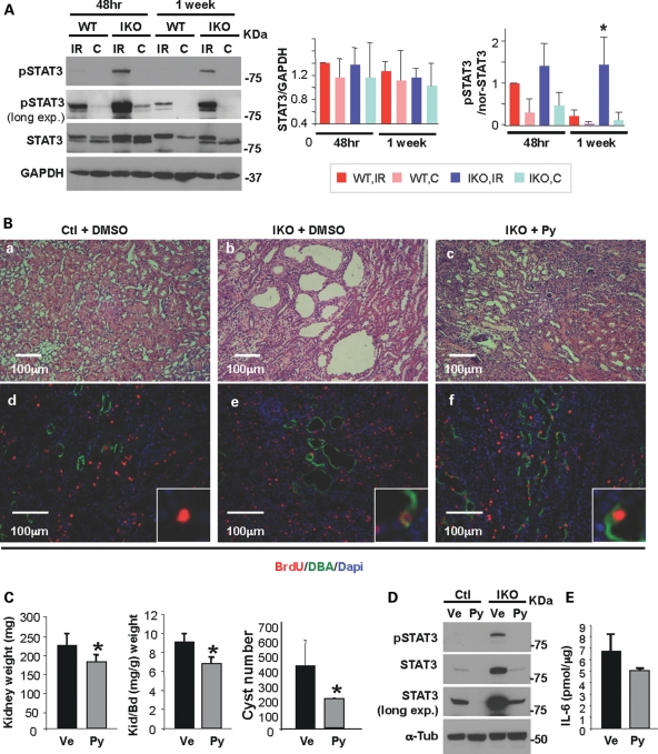
Figure 4.
Pyrimethamine suppresses cyst formation and growth in an adult PKD mouse model. (A) Unilateral IRI was performed on the Pkd1 IKO and their littermate wild-type mice, followed by reperfusion for 48 h or 1 week. Western blot analyses of kidney extracts from Pkd1 IKO (IKO) and control (WT) mice were performed. GAPDH was used as a loading control. IR, injured kidney; C, contralateral (non-ischemic) kidney. Data shown are representative from three mice per group. STAT3 expression is normalized to GAPDH and the ratio of pSTAT3/STAT3 is normalized to the loading control GAPDH [pSTAT3/nor-STAT3, 1 week-WT-IR versus 1 week-IKO-IR, *P< 0.02). (B) Representative H&E staining of kidney sections are shown in (a–c). Kidney sections were double-labeled with anti-BrdU antibody (red) and DBA (green) (d–f). The Pkd1 IKO mice shown here are male Mx1Cre+Pkd1flox/flox mice. (C) Kidney weight (left, *P< 0.05), kidney and body weight ratio (middle, *P< 0.02) and cyst number (right, *P< 0.038) of vehicle- or pyrimethamine-treated Pkd1 IKO mice are shown (n= 4 mice per group). (D) Western blot analyses of kidney extracts from either vehicle- or drug-treated Pkd1 IKO and control mice show that activated STAT3 decreases in both Pkd1 IKO and control kidneys with drug treatment. α-Tubulin was used as a loading control. (E) IL-6 production in kidney lysates from either vehicle- or drug-treated Pkd1 IKO mice was measured by enzyme-linked immunosorbent assay (n= 3 mice per each genotype). The genotype for the Pkd1 IKO mice is Mx1Cre+Pkd1flox/flox. All data shown here were expressed as the mean ± SD. The size of STAT3α, STAT3β, GAPDH and α-tubulin is 79, 86, 37 and 55 kDa, respectively. The protein markers are indicated.

