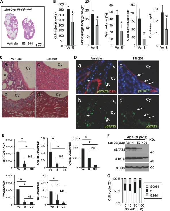
Figure 5.
Selective chemical probe inhibitor of STAT3 reduces cyst formation and growth in a neonatal PKD mouse model. (A) Representative H&E stained whole kidney sections from vehicle-treated (left) and S3I-201 (right)-treated Pkd1 IKO (female Mx1Cre+Pkd1flox/null) mice. (B) Kidney weight (*P< 0.021), kidney and body weight ratio (*P< 0.017), cyst volume (*P< 0.019) and cyst number (*P< 0.025) of vehicle- or S3I-201-treated Pkd1 IKO mice were quantified (n = 4 mice per group). Data are expressed as the mean ± SD. (C) Representative H&E (a and c) and Masson's trichrome (b and d) stained kidney sections from vehicle- (a and b) and S3I-201 (c and d)-treated Pkd1 IKO mice are shown at higher magnification. Collagen deposition is shown in blue. (D) Representative immunostaining images of kidney sections from vehicle- (a and b) and S3I-201 (c and d)-treated Pkd1 IKO mice with pSTAT3 and DBA. Arrows and arrowheads in (a) and (b) point to pSTAT3-positive nuclei of cyst-lining epithelia and interstitial cells, respectively. Arrows in (c) and (d) point to pSTAT3-negative nuclei. Cy represents cyst lumen. (E) Real-time RT–PCR was performed on cDNA from vehicle (Ve)- and S3I-201 (S)-treated Pkd1 IKO kidneys. There was a significant reduction in the expression of STAT3 (P< 0.02) and STAT3 target genes c-Myc (P< 0.008), cyclin D1 (P< 0.03), cyclin D2 (P< 0.04) and bcl-X (P< 0.006) in S3I-201-treated Pkd1 IKO kidneys compared with vehicle-treated ones. Un-treated wild-type kidneys (Ctl) were used to show the baseline expression level of the genes of interest. (F) Immunoblot of pSTAT3 and total STAT3 in ADPKD cells treated with S3I-201 at the indicated concentrations. (G) Flow-activated cytometry analysis of human ADPKD cells treated with either vehicle or S3I-201.

