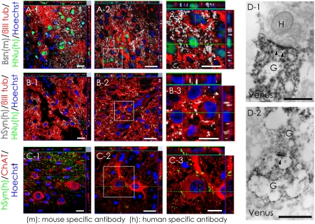
Fig. 2.
Evidence for synapse formation between hiPSC-derived neurons and host mouse spinal cord neurons. (A) Sections were triple-stained with HNu (green), βIII tubulin (red), and the presynaptic marker Bassoon (Bsn, white). The Bsn antibody used here recognized the rat and mouse, but not human, protein. (B) Sections triple-stained for HNu (green), βIII tubulin (red), and the human-specific presynaptic marker hSyn (white). (C) Confocal images showing a large number of somatic and dendritic terminals from graft-derived nerve cells on host motor neurons at the ventral horns. (D) Electron microscopy showing synapse formation between host mouse neurons and graft-derived Venus+ (black) human neurons: the pre- and postsynaptic structures indicated transmission from a host neuron to a graft-derived neuron (D-1) and from a graft-derived neuron to a graft-derived neuron (D-2). H, host neuron; G, graft-derived neuron; arrowheads, postsynaptic density. (Scale bars, 20 μm in A-1, A-2, B-1, B-2, C-1, and C-2; 10 μm in A-3, B-3, and C-3; and 0.5 μm in D.)

