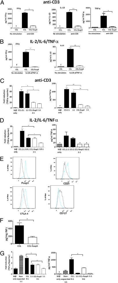
Fig. 4.
Ectopic foxp3 expression attenuates CD4+ effector function. CD4+ lymphocytes isolated from either blood of healthy donors (A–D) or RA synovial MNCs (E–G) were transduced with either pCCL or pCCL-foxp3 lentivirus. NGFR+ cells were restimulated with either anti-CD3 (5 μg/mL) for 24 h or the Tck cytokine mixture for 5 d. RA synovial CD4+ lymphocytes were restimulated with the Tck cytokine mixture. Ratios indicate T cell:macrophage proportions. Representative experiments of >n = 3 are shown and illustrate cytokine production from anti-CD3 activated (A) and cytokine-stimulated (B) T lymphocytes. TNF-α reporter gene and TNF-α protein levels are shown for cocultures with anti-CD3 (C) and cytokine-stimulated (D) T lymphocytes. (E) Phenotype of synovial CD4+ lymphocytes was determined after transduction with pCCL (black) or pCCL-foxp3 (blue) lentivirus. (F) IFN-γ production from RA CD4+ lymphocytes and (G) TNF-α reporter gene activity and protein levels from RA CD4+ lymphocyte:macrophage cocultures after transduction with pCCL-foxp3. *P < 0.05, **P < 0.01, ***P < 0.001, †cytokine concentration below limits of ELISA.

