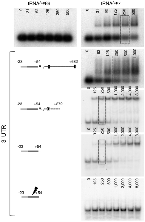
Fig. 3.
Complex formation between tRNAAsp7 and AspRS 3′ UTR. The global organization of the different 3′ UTR variants is displayed. The resolution of the gels was adapted to the length of the tested mRNA: 1% agarose gels for the longest mRNA transcripts (more than 700 nts) and 6% polyacrylamide gels for shorter ones (less than 300 nts). Increasing mRNA concentrations are indicated at the tops of the autoradiograms, and dashed boxes designate the mRNA concentration for which about 50% of the radio-labeled tRNAAsp7 is shifted (Kd). For each variant tested, tRNAAsp69 was used as a control; here only the experiment performed with the full-length mRNA is presented.

