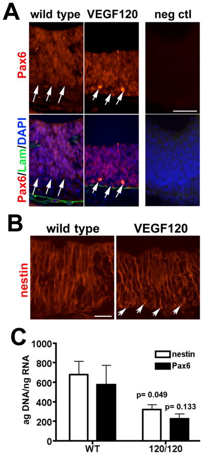
Figure 5. Disruption of VEGF isoform expression alters Pax6 and nestin expression in E11.5 neuroepithelium.

Immunolabeling for (A) Pax6 and (B) nestin is shown in E11.5 neuroepithelium with the ventricular surface at the top and the pial surface at the bottom. (A) The extent of the Pax6-positive domain is indicated by white arrows for wild type and VEGF120 mice and DAPI nuclear stain reveals the cellular population of the neuroepithelium. Labeling for laminin (Lam) in A identifies the limiting basement membrane adjacent to the pial surface and associated with blood vessels. (B) The pattern of nestin filament staining is shown with the white arrows indicating the region of globular nestin expression at the pial surface of VEGF120 embryos. Scale bar is 50 μm in A and 25 μm in B. (C) The qPCR for nestin (white bars) and Pax6 (black bars) are graphed together for comparison, although statistical analyses were conducted separately. RNA from E9.5 wild type and VEGF120 (120/120) neuroepithelium are shown with five or six samples combined from at least three litters. Unpaired t test with Welch’s correction showed that Pax6 expression was reduced by 67% in the VEGF120 compared to wild type, although the observed difference was not significantly different (p = 0.133). Analysis of nestin expression showed a 53% decrease (p = 0.049) in the VEGF120 samples compared to wild type.
