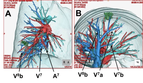
Figure 3.
Images from three-dimensional computed tomographic angiography of the right lower lobe of the lung. The complicated anatomy of the pulmonary arteries (red), pulmonary veins (blue), and bronchus (green) is precisely depicted. From this image, the intersegmental plane between medial basal segment (S7) and anterior basal segment (S8) can be easily imagined.

