
Figure 3.
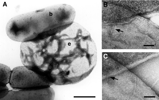
Structural investigation of the bacterium–erythrocyte interaction. (A) After centrifugation the contact between the bacteria (b) and the RBCs (e) was strong enough to withstand water-induced hemolysis. (B and C) Closer inspection showed that the needles of the bacteria pierced the erythrocyte membranes. In both pictures the erythrocyte ghost and the bacterium are in the upper and lower half of the picture, respectively. (The bar is 1 μm for A and 50 nm for B and C.)