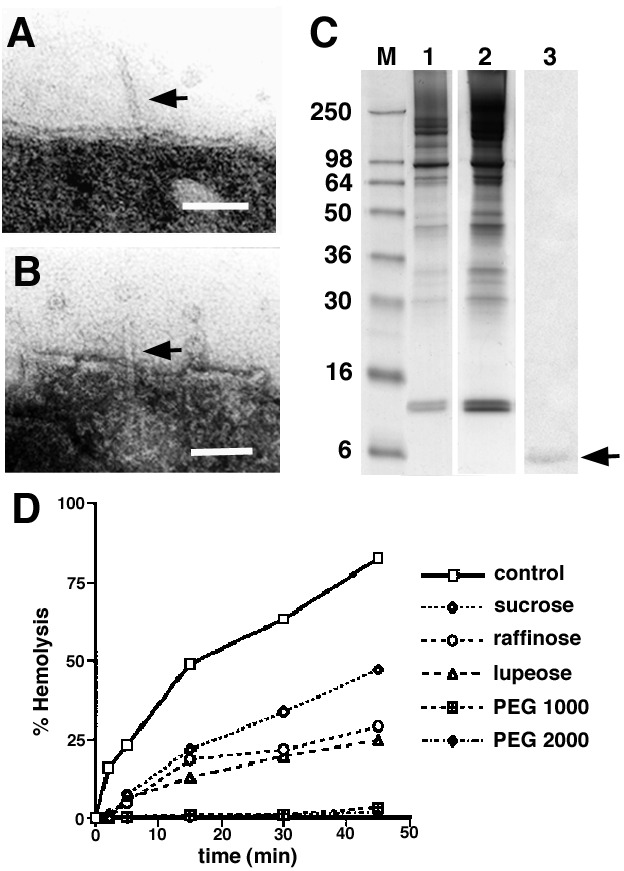
Figure 4.

Examination of the interaction between YscF needles and erythrocyte membranes. (A and B) After separation of bacteria and RBCs, needles are still sometimes found inserted in the RBC membranes. (Bars are 50 nm.) (C) Analysis of the protein pattern of the RBC membrane found before (lane 1) and after (lane 2) contact hemolysis. Bacteria and RBCs were immediately resuspended without incubation at 37°C. Lane 3, immunoblot of lane 2, with an antibody against YscF. (D) Osmoprotection assays were used to determine the size of the bacterially induced lesions. Sugar with a molecular radius of ≥1.8 nm (polyethylene glycol 1000, 2000) blocked the release of hemoglobin and offered osmoprotection.
