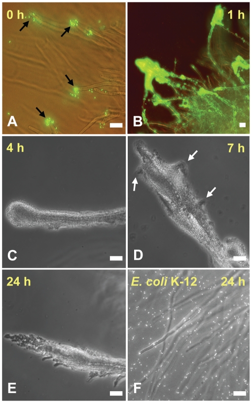
Figure 1. Time course of interaction of S. Typhimurium with A. niger hyphae.
GFP-S. Typhimurium cells attached rapidly to the tip of the fungal hyphae (A, black arrows), where large biofilms formed within 1 h of incubation (B). The biofilm coated the entire hyphae by 4 h (C) and began to differentiate by 7 h (D, white arrows). Distinct branching of the biofilm was visible by 24 h (E). E. coli K-12 (stained with SYTO9®) did not display any significant attachment to the hyphae by 24 h (F). Scale bars, 20 µm.

