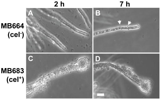
Figure 3. Comparative micrographs of the interaction of the cellulose-deficient S. Typhimurium strain MB664 (cel-, A and B) and its complemented strain MB683 (cel+, C and D), after 2 h (left panel) and 7 h (right panel) of incubation with A. niger.
Strain MB664 failed to attach to the hyphae except for a few rare random cells at later time points (white arrows, B). Transformation with pMB682 restored attachment at high density and formation of a stable biofilm at later incubation times (C and D), albeit not as thick as that of the wild-type strain, as shown in figure 1D. Scale bar, 20 µm.

