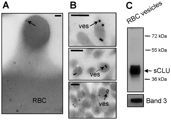
Figure 1. sCLU exocytosis to RBCs-derived vesicles.
(A, B) TEM immunogold localization of sCLU in RBCs membrane protrusions (A) and vesicles (ves) (B) collected from fresh units of stored RBCs (N = 2, young healthy donors). Solid or dashed arrows indicate sCLU immunogold localization at the periphery or the cytosol of the vesicles, respectively. (C) Representative immunoblot analysis of RBCs-derived purified vesicles (N = 2) probed with either polyclonal anti-sCLU or with monoclonal anti-Band 3 antibodies. Molecular weight markers are indicated to the right of the blot. Bars in (A), (B), 100 nm.

