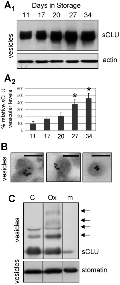
Figure 3. Progressive sCLU accumulation in the RBCs-derived vesicles released during ex vivo aging in blood bank storage conditions.
Representative sCLU immunoblot (A1) and densitometric analysis (A2) of vesicle preparations derived from RBCs units stored in autologous plasma (N = 4). (B) TEM immunogold localization of sCLU at the periphery and the cytosol of vesicles derived from RBCs stored in autologous plasma for 35 days (N = 2). (C) In vitro analysis of sCLU oligomerization pattern (arrows) in vesicles derived from control (C) or tBHP oxidized (Ox) RBCs; m, denotes an isolated plasma membrane sample. Probing with anti-actin and anti-stomatin were used as protein loading references. Shown densitometric data of sCLU vesicular levels (mean values of at least two different experiments) indicate relative proportion against a loading reference followed by normalization against the controls, namely samples stored for a short period of 11 days; error bars indicate ± standard deviation. Asterisks indicate difference of each day of storage vs. day 11 at significance level of p<0.05. Bars in (B), 100 nm.

