
Fig. 3.
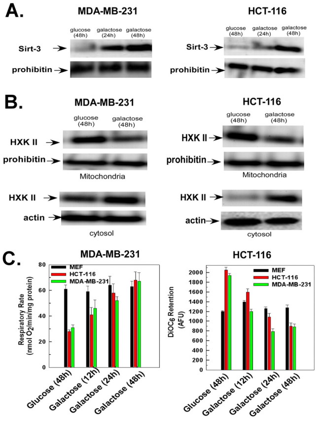
HCT-116 and MDA-MB-231 cells display an increase in sirtuin-3 expression when transferred to galactose-based medium. (A,B) HCT-116 or MDA-MB-231 cells incubated in DMEM containing glucose or galactose for the times indicated. The mitochondrial or cytosolic fractions were used for western blotting and probed with antibodies against hexokinase II or sirtuin-3. Actin and prohibitin were used as loading controls for the cytosolic and mitochondrial fractions, respectively. (C) HCT-116 cells, MDA-MB-231 cells or mouse embryonic fibroblasts (MEFs) incubated in DMEM containing glucose for 48 hours or galactose for the times indicated. Oxygen consumption was measured in a thermostatically controlled chamber equipped with an oxygen electrode. Mitochondrial membrane potential was measured after addition of DiOC6 for the last 30 minutes of incubation. Values are the means from triplicate samples, and the error bars indicate s.d.