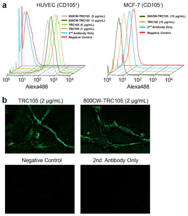
Fig. 1.
In vitro investigation of 800CW-TRC105. a Flow cytometry analysis of TRC105 and 800CW-TRC105 in HUVECs (CD105-positive) and MCF-7 (CD105-negative) cells at different concentrations. b Fluorescence microscopy images of HUVECs using either TRC105 or 800CW-TRC105 (2 μg/mL) as the primary antibody. Various control images are also shown.

