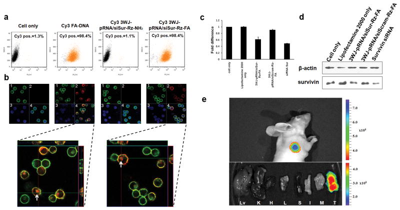
Figure 5. In vitro and in vivo binding and entry of 3WJ-pRNA nanoparticles into targeted cells.
a, Flow cytometry revealed the binding and specific entry of fluorescent-[3WJ-pRNA-siSur-rZ-FA] nanoparticles into folate receptor positive (FA+) cells. Positive and negative controls were Cy3-FA-DNA and Cy3-[3WJ-pRNA-siSur-rZ-NH2] (without FA), respectively. b, Confocal images showed targeting of FA+-KB cells by co-localization (overlap, 4) of cytoplasma (green, 1) and RNA nanoparticles (red, 2) (magnified, bottom panel). Blue–nuclei, 3. c–d, Target gene knock-down effects showed by (c) qRT-PCR with GADPH as endogenous control and by (d) Western blot assay with β–actin as endogenous control. e, 3WJ-pRNA nanoparticles target FA+ tumor xenografts upon systemic administration in nude mice. Upper panel: whole body; Lower panel: organ imaging; Lv=liver; K=kidney; H=heart; L=lung; S=spleen; I=intestine; M=muscle; T=tumor). Scale: Fluorescent Intensity.

