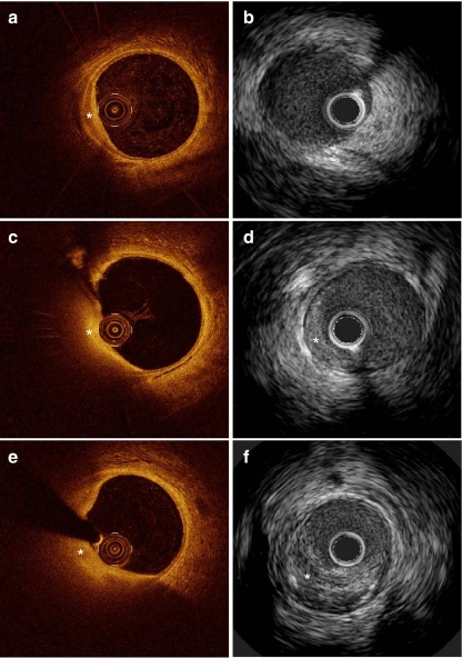
Fig. 1.
Typical example of the development of an atherosclerotic plaque over time on OCT and IVUS in a diabetic pig; a OCT of intimal hyperplasia at 9 months (*); b IVUS of the same cross section as shown in a fails to clearly detect the early lesion; c + d OCT and IVUS of the growing plaque at 12 months (*); e + f OCT and IVUS of the same growing plaque at 15 months (*)

