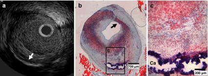
Fig. 2.
Typical example of IVUS (a) and histology (b, c) of a coronary atherosclerotic plaque in a diabetic pig. a IVUS of the same plaque as seen in b and c with deep calcium (white arrow). b and c show an overview and detail of the plaque with circumferential lipid accumulation (stained red) and deep calcification (Ca, remaining rim stained blue). The coronary plaque even shows the presence of a thin fibrous cap (black arrow) overlying the superficial lipid-rich tissue, showing a likeness to a thin cap fibrous atheroma [18]. Oil-red-O stain, bar in b is 700 μm, bar in c is 200 μm

