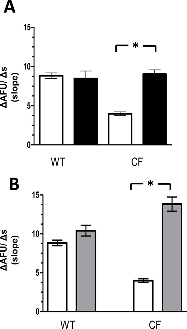
Figure 7. Cl− efflux assessed in low Ca2+ solution.
A): Cl− efflux in macrophages assessed in low Ca2+ solution following carbachol treatment (■) compared with Cl− efflux under control conditions alone (□). (WT= 79 macrophages from 4 mice & CF = 138 macrophages from 4 ΔF508/ ΔF508 and 3 cftr−/− mice). *p<0.0001 B): Cl− efflux in macrophages assessed in low Ca2+ solution following thapsigargin treatment ( ) compared with Cl− efflux under control conditions alone (□). (WT = 27 macrophages from 3 mice & CF = 130 macrophages from 4 ΔF508/ ΔF508 and 4 cftr−/− mice) *p <0.0001 Only CF macrophages demonstrate increases in Cl− efflux following treatment with either carbachol or thapsigargin.

