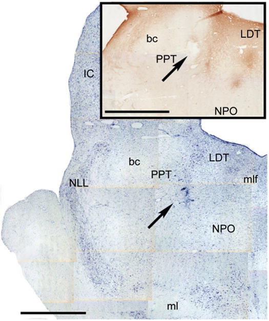
Figure 3.

Microinjetion site of a representative antinsense OD microinjection. The Figure depicts two nearby hemisections counterstained with Nissl and immunostained for NGF (inset) of a representative animal. The arrows indicate the microinjection site. Calibration bars: 2 mm.
