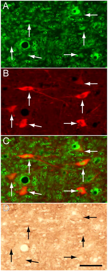
Figure 7.

The antisense OD blocks the production of NGF within the region of injection in the LDT-PPT. An FITC-labeled control antisense was microinjected 18 hours prior to the last antisense OD injection, which preceded euthanasia by approximately 3 hours (see Methods). The FITC-labeled OD was taken up by neurons in the LDT that exhibit immunofluorescent nuclei and cytoplasms (arrows in A). A subpopulation of FITC-labeled neurons are ChAT immunoreactive, i.e., cholinergic in nature (B and the merged image of A and B, in C). The identical field in the LDT shows that these cholinergic cells are devoid of NGF immunoreactivity (D). ChAT immunolabeling was processed with rhodamine; NGF immunoreactivity was visualized with the DAB method. Calibration mark, 40 μm.
