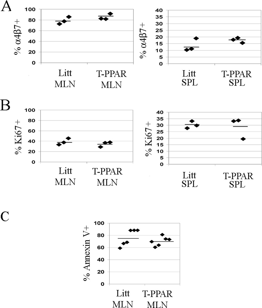
Figure 9. CD8+ T cells from T-PPAR mice do not show abnormalities in expression of α4β7, proliferation, or apoptosis.
Splenic CD8+ cells were purified from littermate or T-PPAR mice by magnetic bead purification. 1 × 10⁶ CD8+ T cells/mouse were injected i.p. into RAG-1−/− mice. MLN and SPL were harvested on day 7 and stained for CD8, MHCII and: A) α4β7; and B) Ki67. MLN were harvested on day 7 and stained for C) CD8 and Annexin V. Results shown are gated on CD8+, MHCII− cells (A,B), or CD8+ cells (C).

