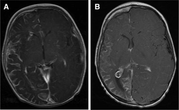
FIGURE 1.

The MRI findings from patient with SWS. A, Contrast-enhanced T1-weighted axial MRI image from patient A obtained at age 5 mos demonstrating right-sided leptomeningeal angioma, consistent with SWS. All lobes of the right hemisphere are noted to be involved. B, Follow-up contrast-enhanced T1-weighted MRI at age 3 additionally reveals atrophy of the entire right hemisphere; a prominent choroid glomus within the right lateral ventricle is also demonstrated on this axial slice. SWS, Sturge-Weber Syndrome; MRI, magnetic resonance imaging.
